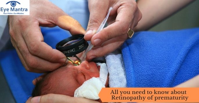
Retinopathy of prematurity (ROP) is an eye-related problem in which there is an abnormal growth of blood vessels near the retina which can result in bleeding inside the eye in premature babies which in severe cases causes loss of vision.
This is also called as Terry syndrome or retrolental fibroplasia (RLF). This is an eye disease seen in prematurely born babies, who have received neonatal intensive care generally. In this neonatal intensive care, oxygen therapy is used for the development of premature lungs.
It is suggested that this condition is caused by disorganized growth of retinal blood vessels, which leads to scarring as well as retinal detachment. It can causes blindness in rare cases but usually gets treated and is mild in nature. It was first discovered by Theodore L Terry in the year 1942. Relative hypoxia and oxygen toxicity contribute to Retinopathy of prematurity development.
Contents
Causes of Retinopathy of prematurity :
During the pregnancy in the fourth month, the retina of the fetus begins its vascularization development. This formation of blood vessels is sensitive to the amount of supplied oxygen, either artificially or naturally. There is a mutation of the NDP gene in some patients who are suffering from said ailment.
Risk factors of Retinopathy of prematurity:
- Cardiovascular problems
- Excessive exposure to oxygen
- Various eye or other infections
- Premature birth or prematurity
- Less birth weight
Pathophysiology:
Generally, the development of retina i.e, growth of blood vessels in center region of retina outwardly. This development of retina gets complete in a few weeks before the delivery of the baby. This is usually seen in normal babies. But, this process is incomplete in premature babies. There is an abnormal growth and branching of blood vessels that causes retinopathy of prematurity condition. These blood vessels grow up from the plane of the retina and can result in bleeding of the retina inside the eye. It can result in detachment of retina and causes loss of vision before 6 months in premature babies.
Retinopathy of prematurity majorly causes fibrovascular proliferation. This fibrovascular proliferation causes the growth of abnormal new blood vessels. Fibrous tissue ( scar tissue) has an association with these newly formed abnormal blood vessels which may result in retinal degradation and detachment of the retina.
There are various multiple factors that influence the progression of the disease such as
- Overall health
- Weight of the child during birth
- Initial diagnosis of the disease
- Stages of retinopathy of prematurity
- Presence or absence of other diseases
The retinopathy of prematurity can be reduced by restricting the supplement oxygen exposure. It is one of the risk factors for development of retinopathy of prematurity condition. By restricting the supplemental oxygen, there is a risk of other hypoxia-related development diseases, including death.
Patients who are suffering retinopathy of prematurity disease can increase the risk of strabismus, cataract surgery ,glaucoma and myopic condition (short sightedness) in later stages of life. So, in order to determine and detect as well as to prevent the other eye conditions, the patient should be examined yearly and given proper treatment.
Prognosis:
Since there are five stages. Stage 1 and stage 2 are mild and do not cause any loss of vision. But the later stages cause an increase in severity of the retinopathy of prematurity condition. Stage 3 of this disease is divided into zone I and zone II based on its effect. Whereas stage 4 and stage 5 cause subtotal and complete retinal detachment respectively.
Few conditions such as squint, amblyopia as well as glaucoma and loss of vision are commonly seen in different stages of retinopathy of prematurity diseases in premature infants.
The retinopathy of prematurity disease is prevalent in developed countries in ranges from 5% to 8% which contains enough neonatal intensive care units and facilities. There is an increased risk of retinopathy of prematurity condition in India, China as well as other Asian countries.
Diagnosis:
The retinopathy of prematurity disease has different stages which are classified by the International classification of Retinopathy of prematurity (ICROP).
The stages are classified as
- Stage 1 is known as the faint demarcation line.
- Stage 2 is known as an elevated ridge.
- Stage 3 is known as extra-retinal fibrovascular tissue.
- Stage 4 is known as sub-total retinal detachment.
- Stage 5 is known as total as well as complete retinal detachment.
Plus disease:
This is one of the major complicating factors at any stage of retinopathy of prematurity condition . This disease is characterized by
- There is a significant level of vascular dilation and curves are observed at the posterior retinal arterioles. This causes increased blood flow through the retina inside the eye.
- Vascular engorgement of iris
- Anterior chamber as well as vitreous haze
- Due to the growth of immature blood vessels over the lens, there is a restriction of pupil dilation. This condition is also called as persistent tunica vasculosa lentis.
Differential diagnosis:
There are other few other diseases that have similarities with retinopathy of prematurity condition such as
- It is a genetic disorder that disturbs the vascularization of the retina in infants. This disease is known as Familial Exudative Vitreoretinopathy.
- Another disease which is known as persistent fetel vasculature syndrome , that causes a traction retinal detachment inside the eye. This one is also called Persistent hyperplastic primary vitreous.
Treatment of retinopathy of prematurity:
In treating retinopathy of prematurity, the peripheral retinal ablation is done majorly. In this treatment, the abnormal or avascular retina is destroyed by performing a solid laser photocoagulation device. This device is easily portable in a neonatal intensive care unit or operating room.
- At first, Cryotherapy is used as the treatment for retinopathy of prematurity disease. It causes destruction of retina by freezing the areas desired. It was found to be an effective treatment as well as for preventing retinopathy of prematurity disease. But due to the availability of laser treatment, there is an decrease in cryotherapy treatment. Cryotherapy treatment has adverse effects such as inflammation and swelling of eyelids.
Surgery:
Vitrectomy surgery or scleral buckling is performed in later stages (stage 4&5) of retinopathy of prematurity disease. It is done in severe cases of retinopathy of prematurity where there is the risk of retinal detachment.
- Intravitreal injections of Avastin (bevacizumab) are used in treating retinopathy of prematurity disease. There are various benefits of this medication, such as a reduced level of anaesthesia usage and the viable peripheral retina is preserved. But there is a risk in the usage of this injection which is ocular and has systemic complications in the patients.
- Propranolol is administered orally for the treatment of the retinopathy of prematurity condition. Propranolol decreases stage 2 progression by almost 48% and stage 3 progress by 52%. However, this medicine causes few adverse effects in infants such as bradycardia and hypotension like conditions.
After treatment:
After the infant is diagnosed and given treatment for retinopathy of prematurity, he or she must be checked yearly for life long. They are needed to perform follow up every year.
After laser or anti-vascular endothelial growth factor treatment, the follow up is individually performed.
Due to the diverse treatments in health care centers of different countries, the check-up of infants (suffering from retinopathy of prematurity disease or not) after the treatment is different in various centers and countries.
The best way to treat your eyes is to visit your eye care professional and get your eyes checked regularly. He will be able to assess the best method of treatment for your eye ailment.Visit our website Eyemantra.To book an appointment call at +91-8851044355. Or mail us at eyemantra1@gmail.com.
Our other services include Retina Surgery, Paediatric Ophthalmologist, Oculoplasty and many more.
Related Articles :


Comments are closed.