The eye is the first amongst our five senses to be treasured. It is also the smallest and the most complex organ in our body. With its maximum diameter just about 2 cms, generally. The anatomy of the eye includes 2 million moving parts. Second only to the brain.
They can distinguish between more than 500 shades of any single color and see more than 2.7 million colors.
How the Eye Works
Sight is all about light. Light reflects from an object in the field of our vision. And it enters our eye through the cornea lens (eye’s front window, the transparent outer covering of the eye). Just behind the thin veil of tears in the front. Passing through this clear layer helps focus the light.
Next is another layer of liquid called the aqueous humor. Its purpose is to circulate throughout the front part of the eye and keep the inside pressure constant. After the aqueous humor, light moves through the iris. This is a colored ring-shaped membrane. It has an adjustable circular opening called the pupil which dilates or compresses to control the amount of light coming in.
Now comes the lens. It operates just like a camera to focus light. It adjusts shape depending on whether the light is reflecting off an object near to you or far away.
The interior part of your eyeball is filled with a gel-like mass called the vitreous humor. After passing through the lens, light has to travel through this humor.
Finally, it hits at the sensitive layer of cells called the retina. At the back, it is “encoded” by a light-sensitive membrane called the retina. The cells are called photoreceptors. The retina transforms the image into electrical messages. as electrical impulses to our brain. Turns them into electrical impulses. These impulses are sent to the optic disk on the retina where they get transferred by a further set of electrical impulses along the optic nerve. And sent to the brain to be processed.
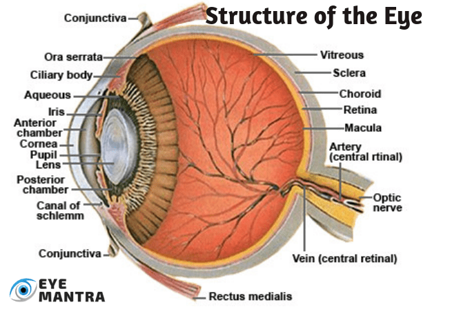
Anatomy of the Eye
Aqueous humor: Aqueous means related to water and humor is fluid. This watery stuff fills the front of the eyeball around the lens.
Blindspot: This is a tiny part of the retina that is not sensitive to light. It is the spot where the optic nerve joins the retina.
Blood vessels: serve to bring oxygen and nutrients to the nerve cells.
Photoreceptors: They are of two kinds: rods and cones. They’re special nerve ends that convert the light into electrochemical signals.
Retinal Pigment Epithelium (RPE): is a layer of dark tissue underneath the Photoreceptors. The purpose of these cells is to absorb excess light so the Photoreceptors can give a clearer signal. They also move nutrients to (and residual or waste from) the Photoreceptors to the Choroid.
Choroid: The Choroid is the middle layer in the anatomy of the eye between the Retina and the Sclera, which is separate from the RPE. It is made up of a lot of fine blood vessels that supply nutrition to the Retina and the RPE. Moreover, it also holds a pigment that absorbs excess light to prevent blurred vision.
Ciliary Body: The Ciliary Body connects the Choroid to the Iris.
Ciliary Muscles: These are tiny muscles surrounding the lens. These muscles hold the lens in place. But they also play an important role in how we see. They squeeze or relax to change the shape of the lens. They squeeze and contract, making the lens fat, to be able to look at nearby objects. And they relax, making the lens thinner, for faraway objects.
Cone cells: It is one of the two types of cells in the Retina, that are light-sensitive. The human Retina contains 6-7 million Cones. They operate best in bright light. They are quite essential for acute vision (receiving a sharp accurate image). The area of the Retina called the Fovea holds the most concentration of cones. Three types of cones are known. Each of them is sensitive to the wavelength of a different primary color – red green or blue. Other colors are seen as a combination of these primary colors.
Conjunctiva: This is the wall on the inside of your eyelid and the outside of the front of your eye (except for the special skin of the Cornea). You can also watch some tiny blood vessels on the Conjunctiva over the anatomy of your eye. Whenever your eyes get sore, these blood vessels get bigger, making your eye looks red.
Cornea: This is the transparent skin that covers the front of your eye. It is clear and slightly convex. This is the see-through part of the eyeball. It has no blood vessels in it.
Fovea: It is a small indentation at the center of the Macula. The Fovea is described as the area with the greatest concentration of Cone cells, the messages encoded at the center of the Fovea will be interpreted by the brain in the form of a visual image.
Iris: Our Iris controls what amount of light would enter our eye. The Iris is the dark-colored part in the anatomy of our eye. It consists of thin circular and longitudinal muscle fibers just behind the cornea. It forms a colored muscular diaphragm across the front of our lens, with an aperture in the center called the Pupil. This expands or contracts to allow more or less light, respectively, depending on the light in the surroundings. The layer of Aqueous Humor prevents the Iris from sticking to the lens behind and the cornea in front.
Lens: The lens is a clear crystalline globe, that focuses light onto the Retina. It almost touches the posterior surface of the pupillary opening. Its shape gets constantly modified to make sure that the ‘picture’ on the retina is as clear as possible. The Ciliary Muscles, attached to the surface of the lens, help the lens to change shape to focus. As the muscles contract, they cause the lens to become more round or long, so that the rays bend more or less, as per requirement. If the object is far away, the Lens needs to bend the light rays from it more sharply, to make them fall on the center of the Retina, where vision is sharpest. For closer objects, the Lens becomes elongated so that light rays are bent less.
Macula: The yellow spot on the Retina at the back of the anatomy of the eye. It surrounds the Fovea, the area that has the greatest concentration of Cone cells, and is, therefore, the area of greatest acuity of vision. When the eye is directed at an object, the part of the image that is focused on the Fovea is the image most accurately registered by the brain. It is situated right at the center of our Retina. Because it’s the focal point of your eye, it has the most special, light-sensitive nerve endings, called Photoreceptors, than any other part.
Optic Disk: The visible portion of the Optic Nerve also found in the Retina. The Optic Disk identifies the start of the Optic Nerves where messages from Cone and Rod cells start from the eye through nerve fibers to the Optic center of the brain. This area is also known as the ‘blind spot’.
Optic Nerve: Optic Nerve is a continuation of the Retina. Starting from the eye at the Optic Disk. It transfers all the visual information it gets to the brain, with the help of millions of nerve fibers branching from the Rods and Cones. It’s similar to the cable that carries all the TV pictures from your aerial to your TV so that you can watch the programs.
Pupil: This is the hole in the center of the Iris. It lets light into our eyes. It shrinks in bright light and expands in dull light.
Retina: The working of the Retina is much the same as a film in a camera. This layer is sensitive to light, lining the interior of the anatomy of our eye. It is made up of light-sensitive cells known as Rods and Cones. The human eye contains approximately 125 million Rods, which are necessary for seeing in dim surroundings. Whereas, Cones function best in bright light. We have about 6 to 7 million of them in the anatomy of our eye. They are indispensable for receiving a sharp accurate image. Cones can also distinguish colors. They turn the picture they receive into an electrical message for the brain, which in turn translates that image to us.
Rod Cells: The other one of the 2 types of light-sensitive cells in the Retina. There are about 125 million Rods, which are crucial for viewing in dim light. They contain a pigment “Rhodopsin” (Visual Purple) which is broken down in the light and regenerated in the dark. Breakdown of the Visual Purple gives rise to nerve impulses when all the pigment is bleached (i.e. in bright light) and the Rods no longer function. This is the condition when the cones get activated.
Sclera: Sclera is the tough skin that covers the outside of the eyeball. Though, it doesn’t cover the transparent Cornea. A protective coat on the “white” part in the anatomy of the eye.
Tear glands: These are small glands inside the upper eyelid. Their purpose is to make tears to keep the surface of the eyeball clean and moist. It helps protect our eyes from damage. When we blink, the tears are spread over the surface of the eye. Tiny particles that may be on our eye (like specks of dust) get washed into the corner of our eyes next to our nose. Sometimes, the tears may flow over our lower eyelids (when we cry or have a fever). But mostly the tears flow down a tiny tube at the edge of your lower eyelid, called the Tear Ducts, next to our nose. The beginning of that tube is visible as a tiny dot. This tube carries the excess tears to the back of our nose. This is why our nose “runs” when we cry.
Vitreous Humor: This is a thicker liquid, gel-like, that fills the larger part of the eyeball and keeps it in shape. Vitreous means glassy, hence the name, because the vitreous humor is very clear so that light can pass through it.
Path of Vision
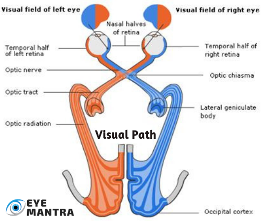
When the light rays fall on the Photoreceptor cells, changes occur in the pigments they contain. This leads to the bleaching of the pigments. And, thus, electrical impulses are generated. These get transmitted through a chain of neurons to the ganglion cells which carry the electrical impulses to the visual cortex of the brain. There they are processed and this is how we see an object.
Each eye receives data from half of the visual field. But the middle parts of both fields overlap slightly. This leads to Binocular Vision. However, the difference in the peripheral portions of the left and right fields of vision leads to the perception of Depth and 3-dimensional vision. It helps in assessing distances accurately and evaluating the depths and dimensions of objects.
Vision Problems/Diseases
The most common problems with vision are Nearsightedness (Myopia), Farsightedness (Hyperopia), Astigmatism – a defect in the anatomy of the eye caused by nonspherical curvature and Age-Related Farsightedness (Presbyopia).
Most people develop Presbyopia in their 40s or 50s and start needing reading glasses. With age, the Lens becomes denser, making it harder for the Ciliary Muscles to bend the Lens.
The leading causes of blindness, in the world, includes Cataracts (clouding of the lens), Age-Related Macular Degeneration (AMD – deterioration of the central retina), Glaucoma (damage to the optic nerve), and Diabetic Retinopathy (damage to retinal blood vessels). Other common disorders are Amblyopia (“Lazy Eye”) and Strabismus (Squint Eyes).
This blog has been shared by EyeMantra, to raise awareness about the eye, its anatomy, various parts, and their functions.
To get your eye checked thoroughly from an expert ophthalmologist, you can make a booking at +91-8851044355. Or mail at eyemantra1@gmail.com.
We also offer various services like Retina Surgery, Specs Removal, Cataract Surgery, and much more.
You may also like: